


Medexplained is an online medical education platform delivering high quality lectures and study materials for medical students and doctors. Medexplained are a team of passionate doctors working diligently to push the envelope regarding post-covid medical education.
‘Black Holes’ is a free 6-month virtual Neuroanatomy lecture series. The course will cover the MRCS neuroanatomy curriculum and also integrate wider content and peer-reviewed journals. They explore anatomy, clinical pathology and key concepts in clinical surgery. Black holes is the first course to carry out augmented reality virtual dissections supplemented with 3D printed anatomical models for VIP members.
Medexplained founder Dr. Jamal Ross , explains that anatomy textbooks do not always give a real understanding of spatial relationships.
What is ‘Black Holes’?
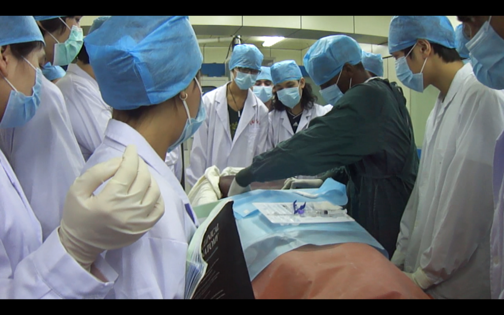
Black holes is a neuroanatomy lecture series and a virtual reconceptualisation of an anatomy course that I taught using cadavers. I distinctly remember the overwhelming smell of formaldehyde, which is commonly used to preserve cadavers for dissections. Formaldehyde is a really pungent gas that can have detrimental effects on the body so I’ve always felt that there must be a safer and better way. The ongoing pandemic has really crystallised some ideas I’ve had around the future of medical education.There are advantages and disadvantages to both scenarios but I believe distance learning is here to stay.
What inspired you?
10 years ago I went on a trip to Sudan and Egypt where I learnt alot about Ancient Nubia and the history of medicine and surgery. I want to teach medical concepts but I also want to tell a story and give some historical context. Its really a blend between a documentary and a lecture.
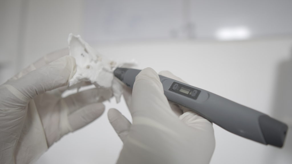
Dr. William Erl Demyer said “ if you can’t draw it, you don’t know it”. This really stuck with me and I started experimenting with drawing structures in 3D with a 3D pen. Its a really huge learning curve, even for someone that knows the anatomy in 2D. Thats when I realised the immense value in what I was doing, because it was so difficult. If you can draw the circle of willis in 3D space then it starts to get really interesting. Most students will only be familiar with these structures in the xy plane because of conventional textbooks.
Understading the tortuousity of the internal carotid artery, for example, and appreciating that in 3 dimensions opens up new ways of learning that translates better to the operative realm.
Can you explain how you use VR and 3D printing ?
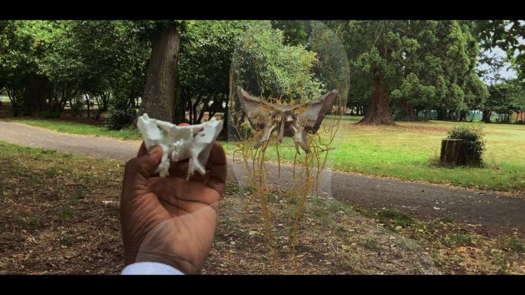
I use a modified VR headset to carry out virtual dissections in serene outdoor environments. It’s really cool being able to do a lecture sitting on a park bench, and simultaneously having a tactile 3D print of the structure you are studying is deeply satisfying: It bridges the virtual to the physical. I’m a huge fan of Netter’s anatomical illustrations but I think this bridge is what is missing from textbooks.
3D printing is such an amazing technology for prototyping and getting ideas into the real world. I started experimenting by printing CT scans and even modifying them to design pathological specimen. In my opinion, it’s one of the most effective ways to communicate with students. Using augmented reality and superimposing that on a physical 3D model creates a kind of ‘Mixed reality’ that is unlike anything i’ve ever tried.
What is next?
If its fun then it’s worth doing. I’m interested in learning more about how people learn. Perhaps some of these lessons will act as a bootloader to something bigger.